
University of South Alabama Mobile
USA Health Adds Robotic Bronchoscopy to Find Hard-to-Reach Lung Cancer Earlier
USA Health University Hospital now offers the Ion robotic bronchoscopy system with AI-assisted CT software to help doctors reach and biopsy small lung nodules and diagnose lung cancer sooner.
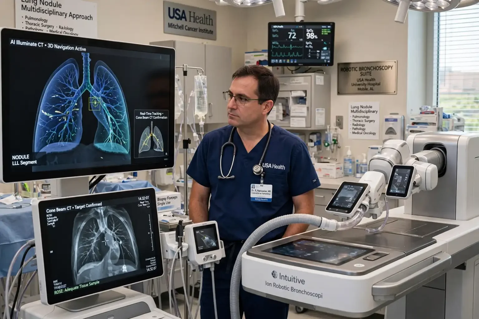
MOBILE, Ala. — USA Health University Hospital has added robotic bronchoscopy technology designed to help doctors find and diagnose lung cancer earlier, giving patients with small or hard-to-reach lung nodules a new option for getting answers close to home.
The hospital now offers the Intuitive Ion Robotic Bronchoscopy system, paired with AI Illuminate CT software and cone beam CT imaging. Together, the tools use 3D mapping, robotic navigation, artificial intelligence, real-time location tracking and robotic stability to guide physicians through the lungs’ branching airways and reach spots that can be difficult to access with conventional bronchoscopy.
“The system positions USA Health as a leader in technology by providing the best equipment within a 100-mile radius,” said Dr. Robert Petrossian, director of Interventional Pulmonology. “This offers patients the best care available for hard-to-reach lung nodules right here at home.”
Why lung nodules matter
Lung cancer is the leading cause of cancer-related deaths worldwide, which makes early detection especially important. It is often found through imaging, including CT scans performed as part of lung cancer screening.
Those scans frequently turn up lung nodules, small spots of tissue that show up on imaging. Most nodules are not cancer. Many are scars from old infections or other harmless growths. But some are early cancers, and the challenge for doctors is telling the difference without putting patients through unnecessary procedures or leaving a dangerous spot unchecked.
Doctors weigh a nodule’s size, shape, growth over time and the patient’s risk factors to decide whether to keep watching it with follow-up scans or take a tissue sample. When a sample is needed, the location of the nodule matters a great deal. Spots deep in the outer parts of the lung, where airways become very narrow, have historically been difficult to reach with a standard bronchoscope, sometimes leading to a needle biopsy through the chest wall or surgery to get a diagnosis.
How the Ion system works
Bronchoscopy is a procedure in which a physician passes a thin tube through the mouth and into the airways of the lungs, usually while the patient is under anesthesia. No incision is made in the chest.
Robotic bronchoscopy adds computer guidance and mechanical precision to that approach. Before the procedure, the patient’s CT scan is used to build a 3D map of the lungs and plan a route through the airways to the nodule. During the procedure, a thin, flexible robotic catheter follows that route while the system tracks its location in real time. Robotic stability holds the catheter steady once it reaches the target, allowing the physician to collect tissue with a biopsy tool.
Cone beam CT, an imaging technique that creates 3D images in the procedure room, can help confirm that the catheter and biopsy tool are in the right spot before samples are taken.
“With the Ion system, we have an advanced way to navigate through the lung and reach small nodules that can be a challenge to access,” Petrossian said. “It allows us to approach these areas with greater control and stability while obtaining tissue samples that can help us make a diagnosis. For patients, this can mean a clearer path from an abnormal scan to understanding what’s causing it and what comes next.”
Answers during the procedure
Once the targeted area is reached, physicians can collect tissue for rapid on-site evaluation, known as ROSE, and for pathology analysis to determine whether a nodule is cancerous.
Rapid on-site evaluation means a pathology specialist examines a quick preparation of the sample while the procedure is still under way. That real-time check helps confirm that enough tissue has been gathered for full pathologic and molecular testing, which reduces the chance a patient will need to come back for a repeat procedure.
Molecular testing has become an important part of lung cancer care. Many lung cancers carry specific genetic changes or markers that can be matched to targeted drugs or immunotherapy, so having enough tissue for those tests can shape treatment decisions.
AI that scans every chest CT
The AI Illuminate CT software adds another layer. According to USA Health, it routinely scans all chest CTs for incidental lung nodules, meaning spots that turn up on scans ordered for other reasons, such as a CT done after an injury or to look at the heart.
Incidental nodules can be overlooked or lost in follow-up because the patient and doctor were focused on another problem. Software that flags those findings can help make sure suspicious spots get a second look. When a suspicious nodule is found, a tissue sample can give physicians the information they need to confirm a diagnosis and decide on the best course of care.
Part of a team approach to lung cancer
Dr. Brian Persing, director of the USA Health Mitchell Cancer Institute and a lung cancer specialist, said the technology will help the hospital’s cancer program move faster and more precisely.
“Adding this technology to our lung cancer program strengthens our ability to provide patients with a timely and accurate diagnosis, which will create more precise treatment options,” Persing said. “This will allow our multidisciplinary team to make informed decisions that will ultimately provide better care and outcomes for our patients.”
Persing said the new tools strengthen USA Health’s comprehensive approach to lung cancer, which brings together specialists in:
- Pulmonology
- Thoracic surgery
- Radiology
- Pathology
- Medical oncology
Those specialists evaluate patients together and develop individualized treatment plans. In a multidisciplinary model, a patient’s scans, biopsy results and overall health are reviewed by several specialties at once, so decisions about surgery, radiation, medication or continued monitoring reflect input from each field.
USA Health is the academic health system of the University of South Alabama, and the Mitchell Cancer Institute is its cancer center. The system has been adding specialized cancer services; earlier this month, it announced the hiring of a fellowship-trained urologic oncologist with expertise in robotic surgery. Read more about that addition.
Who should consider lung cancer screening
Because lung cancer often causes no symptoms in its early stages, screening is one of the best tools for catching it when it is most treatable. The U.S. Preventive Services Task Force recommends annual screening with low-dose CT for adults ages 50 to 80 who have a 20 pack-year smoking history and currently smoke or have quit within the past 15 years. A pack-year equals smoking one pack a day for one year.
People who think they may qualify should talk with their primary care provider, who can review their history, discuss the benefits and risks of screening and place an order if appropriate. Most health insurance plans, including Medicare for eligible beneficiaries, cover lung cancer screening for people who meet the criteria.
Symptoms worth reporting
Anyone with symptoms should see a doctor rather than wait for a screening. Warning signs of lung cancer can include:
- A cough that does not go away or gets worse
- Coughing up blood
- Chest pain
- Shortness of breath or wheezing
- Hoarseness
- Unexplained weight loss or fatigue
What it means for patients
For patients across the Gulf Coast, the practical change is access. Petrossian said the system is the best equipment of its kind within 100 miles, which means people who might otherwise travel farther for a minimally invasive biopsy of a difficult nodule can now have it done in Mobile.
A faster, more accurate diagnosis can shorten the anxious stretch between an abnormal scan and a treatment plan. If a nodule turns out to be benign, patients can avoid more invasive procedures. If it is cancer, the care team can move ahead with tissue already in hand for the testing that guides treatment.